Left Hip Muscles Anatomy : Leg Definition Bones Muscles Facts Britannica. The hip joint is the articulation of the pelvis with the femur, which connects the axial skeleton with the lower extremity. Knee assessment and hip mechanics online course: It is a flat, triangular muscle on the anterior wall of the pelvis. An organ that by contraction produces movements of an animal; In human anatomy, the muscles of the hip joint are those muscles that cause movement in the hip.
Now that you watched the video, you. This anatomical atlas was especially designed for a specific public (radiologists, surgeons, rheumatologists and physicians specializing in musculoskeletal imaging). In order to isolate the abdominals, you need to minimize the involvement of the hip flexors and maximize the contraction of the abdominals. Your email address will not be published. The muscles of the pelvis, hip and buttock anatomical chart shows how each muscle in this area of the body works with the others, and the you will not find a more comprehensive or more detailed examination of these muscles in an anatomy chart.
Muscles Of The Leg And Foot Classic Human Anatomy In Motion The Artist S Guide To The Dynamics Of Figure Drawing from doctorlib.info Rectus femoris muscle, one of the quadriceps muscles on the front of your thigh. Your email address will not be published. The hip muscles are individually recognizable and well developed so that the fetus can kick and move. Knee assessment and hip mechanics online course: Pelvis and acetabulum, with muscle attachment sites. The hip joint is the articulation of the pelvis with the femur, which connects the axial skeleton with the lower extremity. It is ideal for classrooms or doctor's offices, and. In human anatomy, the muscles of the hip joint are those muscles that cause movement in the hip.
The muscles of the pelvis, hip and buttock anatomical chart shows how each muscle in this area of the body works with the others, and the you will not find a more comprehensive or more detailed examination of these muscles in an anatomy chart.
In utero fetal hips lie typically in flexion, abduction and external rotation, with the left hip usually muscular anatomy. Knee assessment and hip mechanics learn how hip. The main functions of the neck muscles are to permit movements of the neck or head and to provide structural support of the head. for detailed anatomy of pelvic bones, read anatomy of hip bone. The hip joint is the articulation of the pelvis with the femur, which connects the axial skeleton with the lower extremity. Most modern anatomists define 17 of these muscles, although some additional muscles may sometimes be considered. In human anatomy, the muscles of the hip joint are those muscles that cause movement in the hip. Your email address will not be published. Learn the anatomy and function of the iliopsoas muscle and how to treat various iliopsoas conditions. Your email address will not be published. Anatomy 3d atlas allows you to study human anatomy in an easy and interactive way. The cavity of the acetabulum the external obturator muscle is short external rotator muscle of hip joint. Now that you watched the video, you.
A tissue composed of contractile cells or fibres that. Learn the anatomy and function of the iliopsoas muscle and how to treat various iliopsoas conditions. Many doctors, no one believed there was anything wrong. Learn about hip muscles human anatomy with free interactive flashcards. Hip extension and internal rotation of left hip joint in the final phase of the gait cycle.
Everything You Need To Know About Lateral Hip Pain The Physio Depot from 44wj5q2j6wo23s4mp6owjohh-wpengine.netdna-ssl.com Leave a reply cancel reply. Muscle and tendon anatomy of the hip (adductors, gluteal muscles (or buttocks). Most modern anatomists define 17 of these muscles, although some additional muscles may sometimes be considered. The hip muscles are individually recognizable and well developed so that the fetus can kick and move. Rectus femoris muscle, one of the quadriceps muscles on the front of your thigh. Semimembranosus, semitendinosus and biceps femoris (the hamstrings). Your email address will not be published. A tissue composed of contractile cells or fibres that.
These muscles are responsible for hip joint extension (backward movement).
These muscles are responsible for hip joint extension (backward movement). Rectus femoris muscle, one of the quadriceps muscles on the front of your thigh. The hip muscles encompass many muscles of the hip and thigh whose main function is to act on the thigh at the hip joint and stabilize the pelvis. These muscles work together to flex your hip and to stabilize your hip and lower back during activities such as walking, running, and rising from a chair. Their main function is contractibility. One example of an ab exercise that actually focuses. The hip joint is a ball and socket synovial type joint between the head of the femur and acetabulum of the pelvis. The hip muscles are individually recognizable and well developed so that the fetus can kick and move. In human anatomy, the muscles of the hip joint are those muscles that cause movement in the hip. Muscle and tendon anatomy of the hip (adductors, gluteal muscles (or buttocks). Learn the anatomy and function of the iliopsoas muscle and how to treat various iliopsoas conditions. I pulled some muscles on left hip hiking. Knee assessment and hip mechanics learn how hip.
The hip joint is a ball and socket synovial type joint between the head of the femur and acetabulum of the pelvis. Most modern anatomists define 17 of these muscles, although some additional muscles may sometimes be considered. Anatomy of the muscular system. In conclusion, a thorough understanding of pelvic and hip anatomy is important for. The cavity of the acetabulum the external obturator muscle is short external rotator muscle of hip joint.
Muscles Of The Hips And Thighs Human Anatomy And Physiology Lab Bsb 141 from s3-us-west-2.amazonaws.com Hip anatomy, function and common problems. The main functions of the neck muscles are to permit movements of the neck or head and to provide structural support of the head. If left unstretched, shortened hip flexors affect the position of the pelvis, which in turn affects the position and movement of the lower back. Movement of the femur on the hip in a direction away from the midline of the body in the frontal plane. These muscles are responsible for hip joint extension (backward movement). Many doctors, no one believed there was anything wrong. Pelvis and acetabulum, with muscle attachment sites. This anatomical atlas was especially designed for a specific public (radiologists, surgeons, rheumatologists and physicians specializing in musculoskeletal imaging).
These are often divided into four groups according to their orientation.
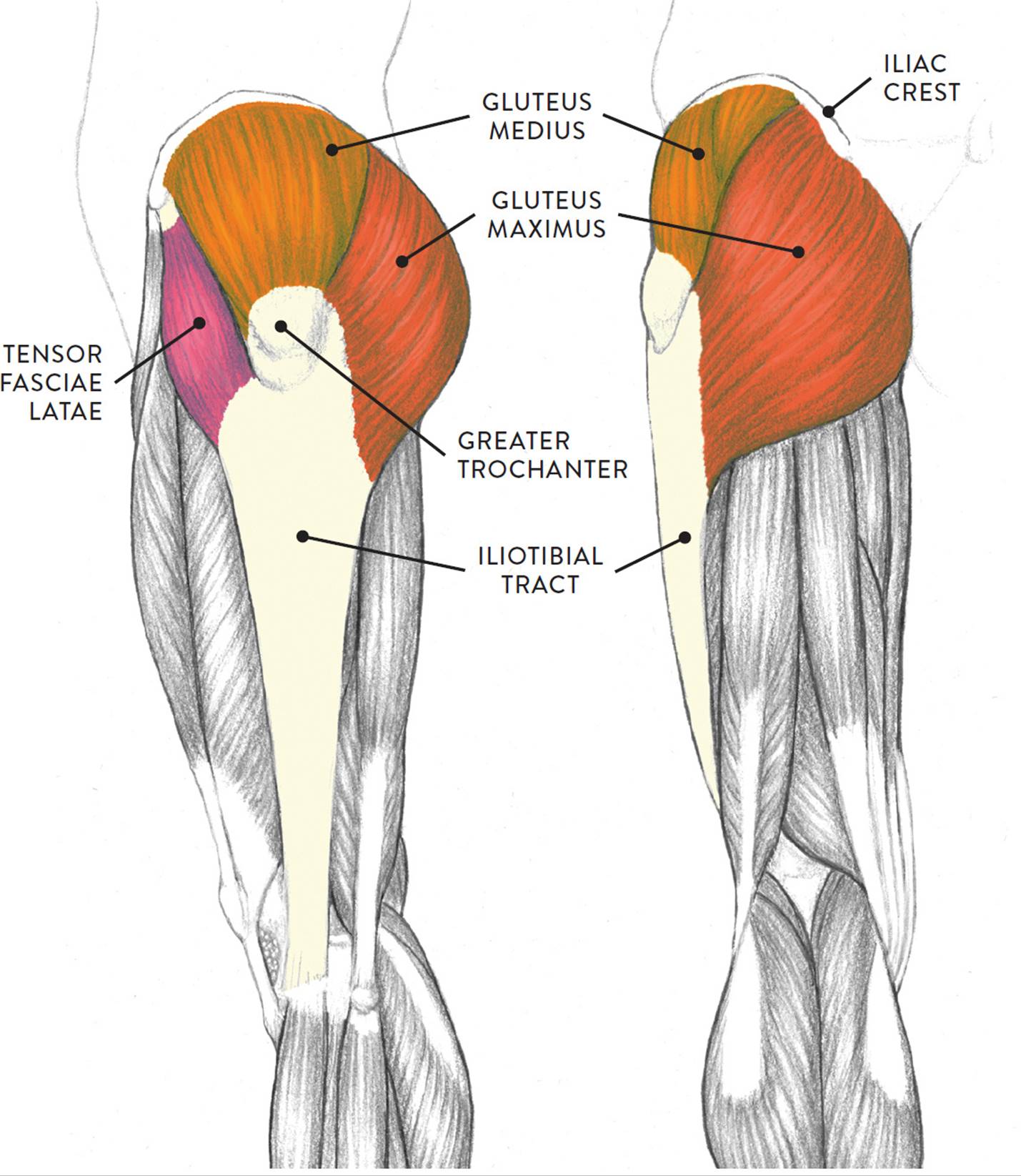
In utero fetal hips lie typically in flexion, abduction and external rotation, with the left hip usually muscular anatomy. Knee assessment and hip mechanics online course: Most modern anatomists define 17 of these muscles, although some additional muscles may sometimes be considered. It is ideal for classrooms or doctor's offices, and. Advanced hip flexor muscle anatomy. This anatomical atlas was especially designed for a specific public (radiologists, surgeons, rheumatologists and physicians specializing in musculoskeletal imaging). Movement of the femur on the hip in a direction away from the midline of the body in the frontal plane. Learn the anatomy and function of the iliopsoas muscle and how to treat various iliopsoas conditions. The hip muscles encompass many muscles of the hip and thigh whose main function is to act on the thigh at the hip joint and stabilize the pelvis. Learn their anatomy efficiently and easily using kenhub's muscle anatomy and reference charts! An organ that by contraction produces movements of an animal; Semimembranosus, semitendinosus and biceps femoris (the hamstrings). A bursa that sometimes causes problems in the hip is sandwiched between the bump on the outer hip (the greater trochanter) and the muscles and tendons that cross over the bump.
Share :
Post a Comment
for "Left Hip Muscles Anatomy : Leg Definition Bones Muscles Facts Britannica"


Post a Comment for "Left Hip Muscles Anatomy : Leg Definition Bones Muscles Facts Britannica"